Cucurbit Genetics Cooperative Report 20:37-40 (article 17) 1997
Zhang Jiannong
Melon Research Institute, Gansu University of Agriculture, Lanzhou, Gansu 730070, P.R. China
The melon mosaic disease described in this note was first observed in Gansu, China, in 1983. The frequency of the disease was less than 1/60,000 plants in a large melon field, but was found in 66.7% of the plants in one breeding line in our experimental plots. The symptoms were different from those of other mosaic diseases caused by cucumber mosaic virus (CMV), watermelon mosaic virus, and other viruses. In 1986-1988, isolation cultures and filtrates of diseased tissue were screened microscopically, and no fungi, bacteria or nematodes were observed. Neither rubbing the inoculum on plants nor aphids spread the disease. Transmission appears to be only through seeds of diseased plants. Previously, seeds from diseased plants were treated with hot water (60 C, 10 min), AgNO 3 (1%), Na 2PO 4 (10%), mercuric chloride (0.1%), and high temperature (73C, 96 hr), but did not cause remarkable differences in the rate of disease (4). We are using this strain to study the expression characteristics and genetics of the disease.
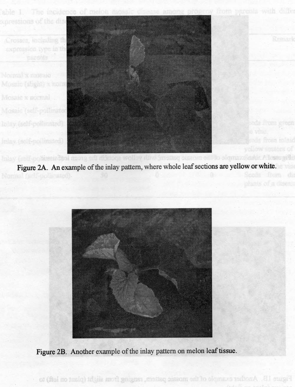
Materials and Methods. Diseased plants were divided into three types based on expression characteristics: (1) “Mosaic,” the most common type (Figures 1A and 1B), with yellow spots of varying sizes in the green tissue of the leaves, and with a clear dividing line between yellow and green. If there were only a few small spots on the leaves, this plant type would grow regularly. If there were many and large spots, or dense small spots, the plants did not grow regularly, sometimes without producing fruits or blossoms. (2) “Inlay” (Figures 2A and 2B), where half or less of leaves were yellow or white, from the leafstalk along the middle or the lateral leaf vein. Sometimes whole leaves or lateral vines were white. Most of these plants could grow regularly, but some were too weak to grow and blossom. (3) “Albino,” where whole cotyledons in seedlings were white, and died about one week after emergence. Albinos arose from seeds with white lateral vines.
In the experimental field, plants with the different types of mosaic disease expression were self-crossed, as well as crossed to normal (cv. Huanghe Honeydew) plants, reciprocally. Seeds were planted individually the following year.
Results and Discussion. The results are presented in Table 1. There was no transmission of the disease when the male parent alone was affected by the symptoms. Among the progeny, the disease symptoms were more serious and the incidence of diseased plants greater when the female parent was affected by the melon mosaic disease. Diseased plants were not found among progeny when the maternal parent was symptomless. Mosaic progeny were observed when the maternal parent expressed either the mosaic or inlay types of the disease. It is postulated that this melon mosaic disease is caused by a plasmagene.
Table 1. The incidence of melon mosaic disease among progeny from parents with different expressions of the disease.
Crossees, including the expression type in the parents |
Number of plants |
Number of diseased plants |
Incidence of disease (%) |
Remarks |
| Normal x mosaic | 103 | 0 | 0 | |
| Mosaic (slight) x normal | 68 | 14 | 20.6 | |
| Mosaic x normal | 51 | 24 | 47.1 | |
| Mosaic (self-pollinated) | 63 | 31 | 49.2 | |
| Inlay (self-pollinated) | 68 | 6 | 8.8 | Seeds from green sectors on the vine. |
| Inlay (self-pollinated) | 51 | 24 | 47.1 | Seeds from inlaid green and yellow sectors of the vine. |
| Inlay (self-pollinated) | 55 | 55 | 100 | Seeds from white (albino) sectors of the vine. |
| Normal (self-pollinated) | 50 | 0 | 0 | Seeds from disease free plants of a diseased strain. |
Figure 1A. An example of the mosaic pattern, with yellow sport in the green leaf tissue.
Figure 1B. Another example of the mosaic pattern, ranging from slight (plant on left) to serious (plant on right).
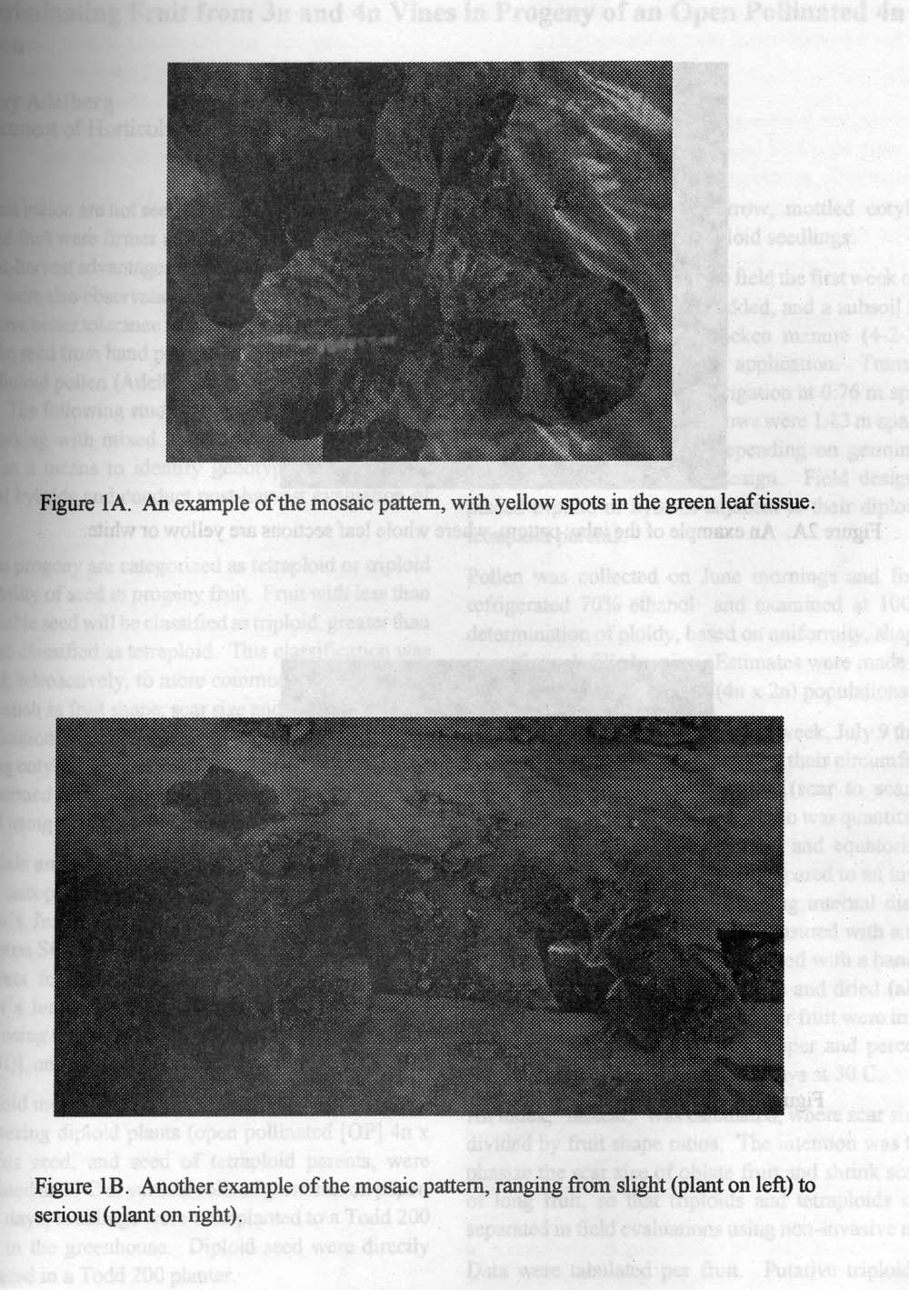
Figure 2A. An example of the inlay pattern, where whole leaf sections are yellow or white.
Figure 2B. Another example of the inlay pattern on melon leaf tissue.
Literature Cited
- Li, G. 1987. Experiment of prevention and cure of hami melon’s viruses. Acta Plant Protect. 14(2):81-85.
- Pitrat, M. 1994. Gene list Cucumis melo L. Cucurbit Genet. Coop. Rpt. 17:135-147.
- Qui, W. 1982. Virology of Plants. p. 427-429.
- Zhang, J.L., An Y. Wei. 1989. A preliminary study on the pathogeny of muskmelon’s inlay mosaic. Acta Agricultural Universitatis Gansu 1:63-68.
- Zhejiang Agricultural University. 1979. Genetics. p. 199-200.
- Zheng, G. 1987. Countermeasure of vegetables viruses. Chinese Vegetables 2:51-54.