Cucurbit Genetics Cooperative Report 22:41-42 (article 15) 1999
Amnon Levi and Claude Thomas
U.S. Vegetable Laboratory, USDA, ARS, 2875 Savannah Highway, Charleston, South Carolina 29414
Our previous experiments in isolating DNA from watermelon leaves using the basic cetyltrimethylammonium bromide (CTAB) procedure (Saghi Maroof et al., 1984; Rogers and Bendich, 1985; Doyle and Doyle, 1987) resulted in: (1) poor DNA yields, (2) co-isolation of highly viscous polysacharides that interfered with DNA handling, (3) co-isolation of polyphenols and other secondary compounds that damaged the DNA by oxidation, and (4) partial or total DNA degradation due to the presence of endogenous nucleases. Thus, we modified the procedure by increasing the CTAB concentration (from 1 to 2.5%) and by adding 0.5% N-lauroyl sarkosine (Sarkosyl) to the DNA extraction buffer, This modification enhanced the breakage of cell and nuclear membranes, and resulted in increased release of DNA into the extraction buffer. It also enhanced the removal of polysaccharides and prevented interaction of polyphenols and endogenous nucleases with the DNA, resulting in high quality as well as high yields of DNA. the procedure presented here has been successfully used in our laboratory.
Procedure:
- Collect young leaves and directly place them in a -80C freezer.
- Grin 5 G leaves to a complete powder using 2 g sand (white quarts, -50 +70 mesh) and three rounds of liquid N.
- Transfer the powder as fast as possible to a 50-ml polypropylene tube. Add 25-ml extraction buffer (60 C). Seal the tube hermetically and shake vigorously to ensure full contact of extraction buffer with tissue. Incubate for 30 min at 60 C. while incubating, shake the tube every 10 min.
- Add 25 ml chloroform and shake the tube vigorously to ensure emulsification of organic and aqueous phases. Open the tube cap to release gas produced by the chloroform. Seal the cap.
- Centrifuge for 5 min at 1,200 relative centrifuge force (RCF) (at 4C). Do not exceed 1,200 RCF, the combination of high detergent concentration, sand and chloroform may crack the tube at higher speeds.
- Place the tubes on ice. Transfer the aqueous phase to a new 50 ml tube, and add 20 ml ice-cold isopropanol. Mix gently but thoroughly, and incubate for 20 min at -20 C.
- Centrifuge for 15 min at 2,200-RCF (at 4 C).
- Drain the tube gently and re-suspend the pellet with 2 ml TE (10:1) containing RNase A (100 μ g/ml). Transfer 1 ml to each of two micro-centrifuge tubes (1.6 ml tube) and incubate for 60 min at 37 C.
- Centrifuge for 5 min at 12,000 rpm in a micro=centrifuge to remove polysaccharides and extraction buffer residues.
- Transfer the supernatant to a new micro-centrifuge tube.
- Add 500 μl chloroform, mix thoroughly, and centrifuge for 5 min at 12,000 rpm (in micro-centrifuge).
- Transfer the upper aqueous phase to a new microcentrifuge tube.
- Add 1/10 5M NaCl (100 μl to 1 ml) and 400 μl isopropanol. Incubate for 20 min at 20 C.
- Centrifuge for 5 min at 12,000 rpm.
- Wash pellet with 1 ml cold ethanol. Drain the tube on a paper towel for about 20 min. (At this stage be careful not to lose the DNA while flipping the tube). Re-suspend in 200-500 μl TE (10:1).
- To clarify, the DNA solution from any residues, centrifuge for 5 min at 12,000 rpm, then transfer the supernatant to a new tube.
Extraction solution: 2.5% CTAB, 0.5% N-lauyrol sarkosine (Sarkosyl), 1.4 M NaCl 100 mM Tris (pH 8.5), 20 mM EDTA. Before extraction add: 1% soluble PVP (average molecular weight 40,000) and 1% insoluble PVP. Also, 5 μl of beta-mercaptoethanol to each 1 ml of extraction solution.
Conclusions
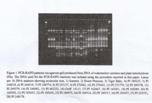
The high concentrations of CTAB and Sarkosyl in the extraction buffer significantly enhanced the quality and yield of DNA isolated from watermelon and melon leaves. Using this procedure we were ale to isolate DNA from a large number of watermelon varieties and plant introductions (PIs), used for PCR-RAPD analysis (Figure 1). The present procedure may also be effective in the isolation of high quality DNA form other cucurbit species.

Acknowledgment: We thank Susan Fox for technical assistance in DNA isolation and in preparing the figure for this manuscript.
Literature Cited
- Doyle, J.J. and J.L. Doyle. 1987. A rapid DNA isolation procedure for small quantities of fresh leaf tissue. Phytochem. Bull. 19:11-15.
- Rogers, S.O. and A.J. Bendich. 1985. Extraction of DNA from milligram amounts of fresh, herbarium and mummified plant tissue. Plant Mol. Biol. 5:69-76.
- Saghai-Maroof, M.A., K.M. Soliman, R.A. Jorgensen, and R.W. Allard. 1984. Ribosomal DNA spacer-length polymorphisms in barley: Mendelian inheritance, chromosomal location and population dynamics. Proc.Natl. Acad. Sci. USA. 81:8014-8018.