Cucurbit Genetics Cooperative Report 18:62-64 (article 29) 1995
T. A. Zitter and J. L. Drennan
Department of Plant Pathology, Cornell University, Ithica, NY 14853
Many cucurbit fruit are susceptible to infection by the black rot fungus Didymella bryoniae in the field or during storage (1, 2, 3). Wounding is known to be important for fungal entry during harvesting and storage operations, but little is known about the impact of rind maturity on infection with and without wounding.
Butternut (Cucurbita moschata cv. Waltham) fruit were collected from the field with the following visual classification: I – pale green, immature; II = beige color; III = tan color; and IV = orange, mature. They were held in a cold room 15C) until inoculated. Squash were gently washed in warm water, air dried, and measured with a Minolta colorimeter which was calibrated for measuring white light. The colorimeter categorized color into three classifications: L value measured lightness; A value distinguished between green and red; and B value distinguished between yellow and blue. There was little variation for the L and B values between fruit, while the A value varied greatly. Fruit were split in half and both halves were placed in plastic boxes lined with moistened paper towels. Fruit were wiped with 70% ethanol and punctured with a 4 mm cork borer. Inoculum consisted of a plug of the fungus cut with a 4 mm cork borer from an actively growing culture of D. bryoniae (Onondaga isolate). Inoculum was placed on two different sites on each fruit half (neck and base), with the wounded and nonwounded sites on the opposite side of each fruit half (ground vs sky side). At each site the fungal plug was covered with a 5 X 5 cm square of clear plastic tape. Boxes were covered and incubated at room temperature. Each inoculation site was measured lengthwise and crosswise at 4 and 7 days after inoculation (DAI). The site wounded with the cork borer had a general wound area of 16, and this value was subtracted from their values in order to provide a more accurate comparison to the nonwounded sites. The experiment was repeated four times during the course of the season and data were analyzed by ANOVA.
There was a strong correlation between the visual categories and the colorimeter values, and suggested that the higher the A value, the less fruit infection would occur. The visual class and corresponding mean A value were I (-5.18), II (0.16), III (2.88), and IV (5.29), respectively, and were correlated at 0.93 (Spearman’s correlation co-efficient). There was no significant difference between the fruit halves (ground vs sky) nor between the neck and the base of the fruit for susceptibility. There were significant differences in lesion size between the different color categories when the mean values were combined for both the wounded and nonwounded areas (Fig. 1). Just 4DAI, fruit in category I were significantly more susceptible than fruit in the other 3 categories. By 7 DAI there were significant differences between categories I and II and between these two categories and categories III and IV. When data were analyzed to separate the importance of wounding vs nonwounding, additional information was learned. Wounding the tissue was a major contributing factor for fungal invasion in all maturity classes (Fig. 2A). The area of tissue invaded was relatively the same for reading taken either 4 or 7 DAI. Although the fungus was able to invade the tissue in all categories without wounding, the amount of invasion was significantly higher for immature fruit in category I, and dropped dramatically as the rind matured (Fig. 2B).
Figure 1. Mean lesion area caused by Didymella bryoniae for each visual class at 4 and 7 days after inoculation (DAI). Ratings taken on the same date with the same letter were not significantly different from one another using Fisher’s LSD (P=0.05).
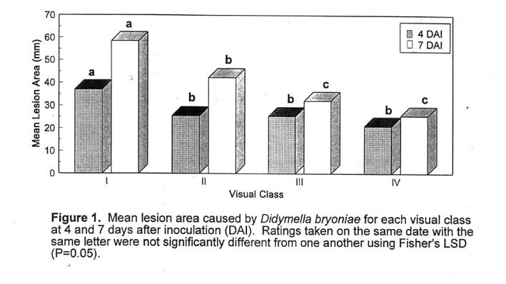
Figure 2. Mean lesion area caused by Didymella bryoniae for (A) wounded and (B) nonwounded tissues for each visual class at 4 and 7 days after inoculation (DAI).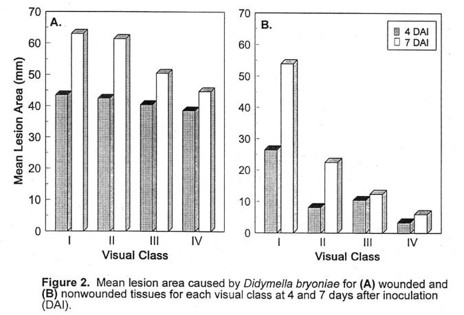
Literature Cited
- deZeeuw, D. J. and H.S. Potter. 1958. PUmpkin black rot in Michigan, 1956. Michigan Agr Expt. Sta. Quart. Bull. 40:477-482.
- Schenck, N.C. 1962. Mycosphaerella fruit rot of watermelon. Phytopathology 52:635-638.
- Steekelenburg, N.A.M. Van. 1982. Factor influencing external fruit rot of cucumber caused by Didymella bryoniae. Neth.J. Pl. Patholo. 88:47-56.